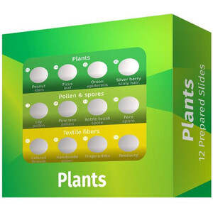
Purtroppo, questa descrizione non è stato tradotto in italiano, in modo da trovare a questo punto una descrizione inglese.
Prepared Microscope Slides
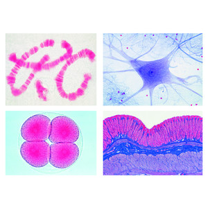
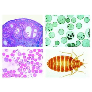
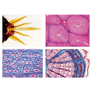
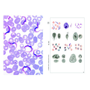
Basic component of the program are the A, B, C and D series comprising of 175 microscope slides. The four series are arranged systematically and constructively compiled, so that each enlarges the subject line of the proceeding one. They contain slides of typical micro-organisms, of cell division and of embryonic developments as well as of tissues and organs of plants, animals and man. Each of the slides has been carefully selected on the basis of its instructional value. LIEDER prepared microscope slides are made in our laboratories under scientific control. They are the product of long experience in all spheres of preparation techniques. Microtome sections are cut by highly skilled staff, cutting technique and thickness of the sections are adjusted to the objects. Out of the large number of staining techniques we select those ensuring a clear and distinct differentiation of the important structures combined with best permanency of the staining. Generally, these are complicated multicolor stainings. LIEDER prepared microscope slides are delivered on best glasses with ground edges of the size 26 x 76 mm (1 x 3"). – Every prepared microscope slide is unique and individually crafted by our well-trained technicians under rigorous scientific control. We therefore wish to point out thatdelivered products may differ from the pictures in this catalog due to natural variation of the basic raw materials and applied preparation and staining methods.
The number of series in hand should correspond approximately to the number of microscopes to allow several students to examine the same prepared microscope slides at the same time. For this reason all slides out of the series can be ordered individually also. So, important microscope slides can be supplied for all students.
No. 500 School Set A for General Biology, Elementary Set 25 microscope slides
Zoology
- Amoeba proteus, w.m. showing nucleus and pseudopodia
- Hydra, w.m. extended specimen to show foot, body, mouth, and tentacles
- Lumbricus, earthworm, typical t.s. back of clitellum showing muscular wall, intestine, typhlosole, nephridia etc.
- Daphnia and Cyclops, small crustaceans from fresh water
- Musca domestica, house fly, head and mouth parts (proboscis) w.m.
- Musca domestica, leg with clinging pads (pulvilli)
- Apis mellifica, honey bee, anterior and posterior wing
Histology of Man and Mammals
- Squamous epithelium, isolated cells from human mouth
- Striated muscle, l.s. showing nuclei and striations
- Compact bone, t.s. special stained for cells, lamellae, and canaliculi
- Human scalp, vertical section showing l.s. of hair follicles, sebaceous glands, epidermis
- Human blood smear, stained for red and white corpuscles - Botany, Bacteria and Cryptogams
- Bacteria from mouth, smear Gram stained showing bacilli, cocci, spirilli, spirochaetes
- Diatoms, strewn slide of mixed species
- Spirogyra, vegetative filaments with spiral chloroplasts
- Mucor or Rhizopus, mold, w.m. of mycelium and sporangia
- Moss stem with leaves w.m. -
Botany, Phanerogams
- Ranunculus, buttercup, typical dicot root t.s., central stele
- Zea mays, corn, monocot stem with scattered bundles t.s.
- Helianthus, sunflower, typical herbaceous dicot stem t.s.
- Syringa, lilac, leaf t.s. showing epidermis, palisade parenchyma, spongy parenchyma, vascular bundles
- Lilium, lily, anthers with pollen grains and pollen sacs t.s.
- Lilium, ovary t.s. showing arrangement of ovules
- Allium cepa, onion, w.m. of epidermis shows simple plant cells with cell walls, nuclei, and cytoplasm -
- Allium cepa, l.s. of root tips showing cell divisions (mitosis) in all stages, carefully stained

